 |
| Image credit: Pexels |
Authors: Ahmed Mamdouh Taha Mostafa, MD; Kevin C. Welch, DO; and Max Cooper, MD RDMS
January/February 2021
Case
A 76-year-old female with a past medical history of hypertension, obstructive sleep apnea, diverticulitis, fibromyalgia, osteoarthritis, depression, and renal cell carcinoma status post remote nephrectomy who presented to our ED with four days of intermittent, diffuse, crampy abdominal pain associated with nausea and non-bloody, non-bilious emesis, hiccoughs, and inability to tolerate PO.
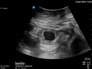
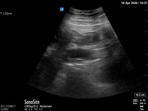
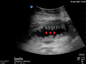
On examination, vital signs were temperature of 98.3º F, pulse of 108 bpm, respiratory rate of 15, blood pressure 146/91 and oxygen saturation of 97% on room air. Significant findings on examination were mild, diffuse tenderness over the abdomen on palpation, which was soft, positive for bowel sounds on auscultation. Bedside ultrasound performed showed keyboard sign – plicae circularis on the interior aspect of the jejunal wall, “to-and-fro” motion, and dilated bowel loops raising suspicion for small bowel obstruction (SBO), which was confirmed by CT.
Laboratory investigations included: white blood cell count 18.3 10*3/uL, hemoglobin 15.7 g/dL, platelets 440,000 10*3/uL, sodium 134 mmol/L, potassium 3.8 mmol/L, chloride 83 mmol/L, bicarbonate 34 mmol/L, BUN 52 mg/dL, creatinine 3.3 mg/dL, glucose 139 mg/dL, lactic acid 1.8 mmol/L, alkaline phosphatase 126 U/L, AST 20 U/L, ALT 13 U/L, and total bilirubin 1.2 mg/dL.
The patient was made NPO, treated with 1 liter of normal saline, morphine and ondansetron, a nasogastric tube was placed, and the surgical team was consulted. The patient was admitted for IV fluids and bowel rest. They were discharged after an uncomplicated hospital course following conservative management.
Discussion
Small bowel obstruction may account for 2% of all ED abdominal pain presentations and may contribute to 300,000 admissions in the United States annually with high rates of severe complications. It represents an important disease entity for consideration in patients with abdominal complaints. According to one study, the best predictors of SBO on history and physical examination were previous abdominal surgery, constipation, abnormal bowel sounds, and/or abdominal distention.1 CT is not only the gold standard for diagnosis of SBO but it also plays an important role in delineating the etiology and, therefore, in operative planning.2 However, it is important to note that there is a relationship between early diagnosis and the decreased requirement for surgical intervention.3 Despite this significance, CT is not always readily available in many settings such as low resource hospitals, multiple simultaneous high priority patients (e.g. CVA, trauma), technical difficulties (machine malfunction, difficult transport). That combined with the fact that multiple studies have reported that bedside ultrasound has comparable sensitivity and specificity to CT, point us to consider it as an important adjunct or alternative in the diagnosis of SBO.1,2,4-6 In addition to being a less costly, more rapid test, that providers can be easily trained in, ultrasound has not been associated with increased risk of cancer due to radiation. It also allows for serial examination to assess for resolution.6
Signs of bowel obstruction on ultrasound include:
- Dilated bowel loops with most studies using >25 mm as the cut-off for diagnosis (Figures 1 and 2).
- “Tanga” sign: free fluid between loops taking a “pointy” triangular appearance, hence the name after the bikini bottom.
- ‘To-and-fro’ motion: hyperechoic bowel contents moving back and forth within the bowel lumen.
- “Keyboard” sign: visualization of the plicae circularis (Figure 3).
References
- Taylor MR, Lalani N. Adult small bowel obstruction. Acad Emerg Med. 2013;20(6):528-544. doi:10.1111/acem.12150
- Frasure SE, Hildreth AF, Seethala R, Kimberly HH. Accuracy of abdominal ultrasound for the diagnosis of small bowel obstruction in the emergency department. World J Emerg Med. 2018;9(4):267-271. doi:10.5847/wjem.j.1920-8642.2018.04.005
- Bickell N, Federman A, Aufses A. Influence of time on risk of bowel resection in complete small bowel obstruction. J Am Coll Surg. 2005;201(6):847-854.
- Jang T, Schindler D, Kaji A. Bedside ultrasonography for the detection of small bowel obstruction in the emergency department. Emerg Med J. 2011;28(8):676-678.
- Unlüer E, Yavaşi O, Eroğlu O, Yilmaz C, Akarca F. Ultrasonography by emergency medicine and radiology residents for the diagnosis of small bowel obstruction. Eur J Emerg Med. 2010;17(5):260-264.
- Gottlieb M, Peksa GD, Pandurangadu AV, Nakitende D, Takhar S, Seethala RR. Utilization of ultrasound for the evaluation of small bowel obstruction: A systematic review and meta-analysis. Am J Emerg Med. 2018;36(2):234-242. doi:10.1016/j.ajem.2017.07.085


