 |
| This post was peer reviewed. Click to learn more. |
Author: Eric Sulava, MD
Emergency Medicine Resident
Naval Medical Center Portsmouth
AAEM Education Committee
Author: Katrina Destree, MD
Staff Physician
Naval Medical Center Camp Lejeune
I am a military service member. This work was prepared as part of my official duties. Title 17 U.S.C. 105 provides that “Copyright protection under this title is not available for any work of the United States Government.” Title 17 U.S.C. 101 defines a United States Government work as a work prepared by a military service member or employee of the United States Government as part of that person’s official duties.
The views expressed in this article are those of the author(s) and do not necessarily reflect the official policy or position of the Department of the Navy, Department of Defense, or the United States Government.
Patient Presentation
An otherwise healthy, 7-year-old boy presents to the emergency department (ED) complaining of right elbow pain following a three-foot fall. The patient states that he was standing on an elevated cable box when he fell, landing directly on his flexed elbow. He complains of diffuse pain and inability to extend his arm. He denies any head/neck injury, loss of consciousness, other injuries, numbness or tingling.
On exam, the patient is well appearing with normal vital signs. Primary trauma evaluation is benign, with no neurologic, cardiovascular, respiratory, abdominal, or pelvic abnormalities. On extremity exam, the patient has right elbow tenderness and swelling surrounding the radial head, olecranon and distal humerus. The swelling extends across the joint line into the proximal forearm. He is neurovascularly intact distally, with full range of motion, sensation and pulses in the fingers.
Questions (see answers below)
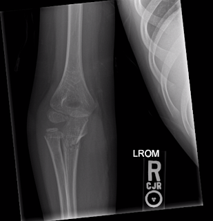
- What is the injury shown in the above radiographs?
- What findings would suggest the need for operative fixation in the above fractures?
Management
Due to the patient’s high-impact injury, limited range of motion, and prominent swelling, radiographs were immediately taken. Although the images were somewhat limited due to the patient’s pain, a transverse fracture of the proximal ulnar metaphysis as well as a non-displaced radial neck fracture were still identified. An orthopedic surgeon was consulted, and the rare presentation of a simultaneous proximal radius and ulna fracture was discussed. The fractures were nondisplaced and minimally angulated, so orthopedics proceeded with closed reduction and casting. The patient was placed in a posterior long arm splint (elbow at 90 degrees with forearm maintained in a neutral position), with an overlying sugar tong splint. The patient followed up withs orthopedic two days later and the splint was exchanged for a long arm cast. The patient remained neurovascularly intact with soft compartments and full distal range of motion. On post-injury day ten, repeat radiographs were taken, showing stable healing of the fractures with appropriate alignment and joint spacing. The patient had no complications during his course of care.
Discussion
Forearm fractures are the most common fractures in children, representing 40-50% of all childhood fractures.[1,2] A majority of these injuries occur following direct blow or a fall onto an outstretched hand (FOOSH), leading to midshaft or distal radius fractures. Proximal injuries, defined as radial head or neck fracture, or ulnar olecranon or coronoid process fractures, are the least common area of injury.[3] These are rare in isolation, and little medical literature exists on proximal both-bone fractures.
These rare injuries usually occur following a high-impact traumatic event, including football, all-terrain vehicle riding, or skateboarding. Due to their traumatic nature, evaluation in the ED should include a primary survey to exclude life threats and central nervous system (CNS) injuries. Anatomically, the elbow and proximal forearm are very high-risk areas for neurovascular compromise. The brachial artery, median nerve, and radial nerve course anterior to the elbow joint, and are at risk with valgus displacement of proximal forearm injuries. The ulnar nerve courses medially through the elbow and can be compromised with posterior injuries.[4] This highlights the importance of evaluating for pulses, perfusion, and neurologic function.
As mentioned above, disposition depends on the fracture pattern, degree of angulation, and level of displacement. Radial head or neck fractures can tolerate up to thirty degrees of angulation before closed reduction is necessary.[3] If greater than thirty degrees of residual angulation is present following closed reduction, operative care should be considered.[5] Anatomically, the proximal area of the ulna contains the olecranon and proximal ulnar shaft. Surgical intervention should be considered for olecranon fracture if there is displacement, step-off over two millimeters, comminution, or accompanying radial head or neck fracture.[3,6] This case involved an isolated proximal ulnar metaphysis fracture, without the classic “Monteggia” pattern of accompanied radial head dislocation. Isolated ulnar fractures with more than fifty percent displacement, ten degrees of angulation, involvement of the proximal third, or distal radioulnar joint instability are considered unstable.[7] In the pediatric population, surgical management should be considered in ulnar fractures with associated severe soft tissue injury, compartment syndrome, or distal humerus fractures.[8]
With exception to the above-mentioned cases, these fractures can be acutely managed with a posterior long arm splint or cast and adequate pain control. Neurovascular status and compartment checks should be completed before and after splinting. Some loss of supination and pronation can be expected with a proximal radial fracture. The rare and serious complications, such as avascular necrosis, growth arrest, elbow deformity, contracture, and nonunion are associated with the following risk factors: age over ten years, angulation over thirty degrees, delayed reduction, and multiple injuries.[3,5,9] Therefore, prompt identification, treatment, and consultation should be completed with these high-impact injuries.
Pearls
- Emergent orthopedic consultation should be considered with: open fractures, neurovascular compromise, forearm fracture with joint dislocation, and acute compartment syndrome.
- Angulation greater than thirty degrees in a radial head fracture justifies reduction.
- Surgical intervention should be considered for olecranon fractures with displacement, step-off over two millimeters, comminution, or accompanying radial head or neck fracture.
Answers
- Transverse fracture of the proximal ulnar metaphysis with a radial neck fracture
- Indications for operational fixation of radial neck fractures: Greater than thirty degrees of angulation, loss of pronation or supination (less than forty-five degrees of mobility)
Indications for operational fixation of proximal ulnar fractures: proximal third ulnar fractures in isolation can be considered unstable due to poor functional result with non-operative treatment [7]
Resources:
1. Price CT, Flynn JM. Management of fractures. Lovell and Winter’s Pediatric Orthopaedics. Philadelphia, PA. Lippincott Williams & Wilkins; 2013.
2. Rodríguez-Merchán EC. Pediatric fractures of the forearm. Clin Orthop Relat Res. 2005;(432):65-72.
3. Lins RE, Simovitch RW, Waters PM. Pediatric elbow trauma. Orthop Clin North Am. 1999;30(1):119-32
4. Rang M, Pring ME, Wenger DR. Rang’s Children’s Fractures. Philadelphia, PA. Lippincott Williams & Wilkins; 2005.
5. Steinberg EL, Golomb D, Salama R, Wientroub S. Radial head and neck fractures in children. J Pediatr Orthop. 1988;8(1):35-40.
6. Gicquel PH, De billy B, Karger CS, Clavert JM. Olecranon fractures in 26 children with mean follow-up of 59 months. J Pediatr Orthop. 2001;21(2):141-7.
7. Sauder DJ, Athwal GS. Management of isolated ulnar shaft fractures. Hand Clin. 2007;23(2):179-84.
8. Herman MJ, Marshall ST. Forearm fractures in children and adolescents: a practical approach. Hand Clin. 2006;22(1):55-67.
9. Caterini R, Farsetti P, D’arrigo C, Ippolito E. Fractures of the olecranon in children. Long-term follow-up of 39 cases. J Pediatr Orthop B. 2002;11(4):320-8.

